Location & Hours
4008 Red Cedar Dr D-1
Highlands Ranch, CO 80126-8152
| Mon & Fri: | 8 - 4 |
| Tues - Thurs: | 10 - 7 |


Do you have floaters in your vision?
Floaters are caused by thick areas in the gel-like fluid that fills the back cavity of your eye, called the vitreous.
Many people, especially highly near-sighted people, often see some number of floaters for a good portion of their lives. Often, these floaters are in the periphery of the vision and may only be visible in certain lighting conditions. The most frequent conditions are when in bright sunlight looking toward the clear blue sky. I know this from personal experience since I have a floater in my left eye that I most often see when swimming outdoors. Every time I turn my head to the left to breathe I see this floater moving in my peripheral vision.
This is totally harmless other than when I’m swimming in the ocean and swear that sudden object in my peripheral vision is a shark bearing down on me. Some people who have floaters are not as lucky-- the floater might be in their central vision and is almost constantly annoying, especially when trying to read.
The second scenario in which floaters occur is during the normal aging process. The vitreous gel in the back of the eye starts to shrink as we age and at some point it collapses in on itself and pulls away from the retina. This sometimes results in a sudden set of new floaters.
When that happens you need to be checked for signs of a retinal tear or detachment. As long as your retina survives that episode without any problems, the floaters themselves may stick around for a while and can be rather annoying.
Most people eventually adapt to the floaters; the brain learns to filter them out so they are no longer aware of them. The vitreous can also collapse more as time goes on and the dense floater can initially may move further forward and drop lower in the eye so the shadow it is casting is less intense and more in the periphery of your vision where it is much easier to ignore.
The first line of treatment for floaters has been, and still is, to learn to live with them. Once you have your retina checked and verify that there is nothing wrong there, the floaters themselves are harmless and will not lead to any further deterioration of your vision--which is why, if at all possible, you should just live with them. This is especially true if the floaters are new because the overwhelming majority of people with new floaters will eventually get to the point where they are no longer seeing them or at least where they are not interfering with normal daily activities.
If you have tried to wait them out and live with them but they are still interfering with your normal daily activities, you may want to consider having them treated with a laser.
This treatment involves using a special laser to try to break down large floaters into much smaller pieces that may no longer be visible. In a study of the laser treatment involving 52 patients, 36 were treated with the laser (a single laser treatment session) and 16 people had a sham treatment (meaning they went through everything the treated group did but did not actually have the real treatment done). In the people who were actually treated, 54% reported a significant improvement in the floater symptoms while 0% in the sham group reported any improvement (no placebo effect). There were no significant side effects in either group.
Some points to note in the above study:
54% of people treated noted a significant improvement in their floater symptoms with a single treatment. That’s clearly not anywhere near a guaranteed improvement.
Other people have noted an improvement after more than one session, bringing the total expected improvement into the 70% range, with one or more treatments.
Another point to note is that there were no significant side effects to the treatment.
Although true in this small study, it does not mean that there are no risks to the laser treatment. Although rare, there have been reports of damage to the retina, optic nerve, or the lens of the eye.
Another treatment that can be used to treat floaters is a surgical procedure called a vitrectomy. This involves surgically going inside the back of the eye and removing the vitreous. This surgical procedure carries a higher risk than the laser treatment and is not 100% effective.
In summary, laser treatment is a good addition to the tools to deal with significant floater problems. If you have floaters for at least six months and they are in your central vision and interfering with your normal daily activities and you want to see if this laser treatment could be right for you, check with your eye doctor.
Article contributed by Dr. Brian Wnorowski, M.D.
This blog provides general information and discussion about eye health and related subjects. The words and other content provided in this blog, and in any linked materials, are not intended and should not be construed as medical advice. If the reader or any other person has a medical concern, he or she should consult with an appropriately licensed physician. The content of this blog cannot be reproduced or duplicated without the express written consent of Eye IQ.
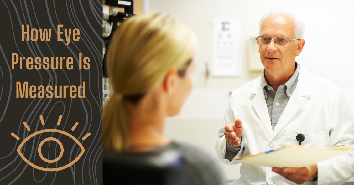
A common question asked during the eye exam is, “When is the puff coming?”
Patients are referring to air-puff or non-contact tonometry. Tonometry is the procedure used to measure eye pressure, and this is important for diagnosing and monitoring glaucoma.
In non-contact tonometry, a puff of air is used to measure the pressure inside the eye. The benefit of this test is there is no actual contact with the eye, but the air puff is sometimes very startling for patients. Some people hate that test and it isn’t the most accurate way to measure your eye pressure.
Some doctors don’t even use the air-puff test. Instead, they place a yellow drop that consists of a numbing medicine and then shine a blue light on the eye. This is done in front of the slit lamp and a small tip gently touches the eye to measure the eye pressure. This procedure is called Goldmann tonometry and is considered the gold standard for measuring eye pressure.
Another method for checking eye pressure is the Tonopen. This is a portable, hand-held instrument that is useful when patients can’t sit in front of the slit lamp to have their eye pressure checked. The Tonopen also requires a numbing drop to be placed in the eye, and the tip gently touches the eye.
A common question related to tonometry is “what is normal eye pressure?”
Normal eye pressure ranges from 10-21 mm Hg. Eye pressure doesn't have any relationship to blood pressure. Many times, people are surprised that their eye pressure is high, but they have normal blood pressure. In general, there is no diet or exercise that will significantly affect eye pressure. It is therefore important to have your eye pressure checked regularly because there are usually no symptoms of high eye pressure until it has affected your vision.
Article contributed by Dr. Jane Pan
This blog provides general information and discussion about eye health and related subjects. The words and other content provided in this blog, and in any linked materials, are not intended and should not be construed as medical advice. If the reader or any other person has a medical concern, he or she should consult with an appropriately licensed physician. The content of this blog cannot be reproduced or duplicated without the express written consent of Eye IQ.
We commonly see patients who come in saying that their eyes are bleeding.
The patient is usually referring to the white part of their eye, which has turned bright red. The conjunctiva is the outermost layer of the eye and contains very fine blood vessels. If one of these blood vessels breaks, then the blood spreads out underneath the conjunctiva. This is called a subconjunctival hemorrhage.
A subconjunctival hemorrhage doesn't cause any eye pain or affect your vision in any way. Most of the time, a subconjunctival hemorrhage is asymptomatic. It is only noticed when looking at the mirror or when someone else notices the redness of the eye. There should not be any discharge or crusting of your lashes. If any of these symptoms are present, then you might have another eye condition that could need treatment.
What causes a subconjunctival hemorrhage? The most common cause is a spontaneous rupture of a blood vessel. Sometimes vigorous coughing, sneezing, or bearing down can break a blood vessel. Eye trauma and eye surgery are other causes of subconjunctival hemorrhage. Aspirin and anticoagulant medication may make patients more susceptible to a subconjunctival hemorrhage but there is usually no need to stop these medications.
There is no treatment needed for subconjunctival hemorrhage. Sometimes there may be mild irritation and artificial tears can be used. The redness usually increases in size during the first 24 hours and then slowly decreases and fades. It often takes one to two weeks for the subconjunctival hemorrhage to be absorbed. The larger the size of the hemorrhage, the longer it takes for it to fade.
Having a subconjunctival hemorrhage may be scary initially, but it will get better in a couple of weeks without any treatment. However, redness in the eye can have other causes, and you should call your eye doctor--particularly if you have discharge from the eye.
Article contributed by Dr. Jane Pan
This blog provides general information and discussion about eye health and related subjects. The words and other content provided in this blog, and in any linked materials, are not intended and should not be construed as medical advice. If the reader or any other person has a medical concern, he or she should consult with an appropriately licensed physician. The content of this blog cannot be reproduced or duplicated without the express written consent of Eye IQ.

Diabetic retinopathy is an eye condition that can affect the retina of people who have diabetes.
The retina is the light-sensitive tissue that lines the back of the eye, and it detects light that is then processed as an image by the brain. Chronically high blood sugar or large fluctuations in blood sugar can damage the blood vessels in the retina. This can result in bleeding in the retina or leakage of fluid.
Diabetic retinopathy can be divided into non-proliferative or proliferative diabetic retinopathy.
Non-proliferative diabetic retinopathy: In the early stage of the disease, there is weakening of the blood vessels in the retina that causes out-pouching called microaneurysms. These microaneurysms can leak fluid into the retina. There can also be yellow deposits called hard exudates present in the retina from leaky vessels.
Diabetic macula edema is when the fluid leaks into the region of the retina called the macula. The macula is important for sharp, central vision needed for reading and driving. The accumulation of fluid in the macula causes blurry vision.
Proliferative diabetic retinopathy: As diabetic retinopathy progresses, new blood vessels grow on the surface of the retina. These blood vessels are fragile, which makes them likely to bleed into the vitreous, which is the clear gel that fills the middle of the eye. Bleeding inside the eye is seen as floaters or spots. Over time, scar tissue can then form on the surface of the retina and contract, leading to a retinal detachment. This is similar to wallpaper contracting and peeling away from the wall. If left untreated, retinal detachment can lead to loss of vision.
Symptoms of diabetic retinopathy:
- Asymptomatic: In the early stages of mild non-proliferative diabetic retinopathy, the person will usually have no visual complaints. Therefore, it is important for people with diabetes to have a comprehensive dilated exam by their eye doctor once a year.
- Floaters: This is usually from bleeding into the vitreous cavity from proliferative diabetic retinopathy.
- Blurred vision: This can be the result of fluid leaking into the retina, causing diabetic macular edema.
Risk factors for diabetic retinopathy:
- Blood sugar. Lower blood sugar will delay the onset and slow the progression of diabetic retinopathy. Chronically high blood sugar and the longer the duration of diabetes, the more likely chance of that person having diabetic retinopathy.
- Medical conditions. People with high blood pressure and high cholesterol are at greater risk for developing diabetic retinopathy.
- Ethnicity. Hispanics, African Americans, and Native Americans are at greater risk for developing diabetic retinopathy.
- Pregnancy. Women with diabetes could have an increased risk of developing diabetic retinopathy during pregnancy. If they already have diabetic retinopathy, it might worsen during pregnancy.
Article contributed by Jane Pan M.D.
This blog provides general information and discussion about eye health and related subjects. The words and other content provided in this blog, and in any linked materials, are not intended and should not be construed as medical advice. If the reader or any other person has a medical concern, he or she should consult with an appropriately licensed physician. The content of this blog cannot be reproduced or duplicated without the express written consent of Eye IQ.

In light of the holiday season, here are our top 10 eye care jokes.
1) What do you call a blind deer? No Eye Deer!
2) What do you call a blind deer with no legs? Still No Eye Deer!
3) Why do eye doctors live long lives? Because they dilate!
4) Why did the blind man fall into the well? He couldn’t see that well.
5) Why shouldn’t you put avocados on your eyes? Because you might get guac-coma!
6) What did the right eye say to the left eye? "Between you and me, something smells."
7) A man goes to his eye doctor and tells the receptionist he’s seeing spots. The receptionist asks if he’s ever seen a doctor. The man replies, “No, just spots.”
8) How many eye doctors does it take to screw in a light bulb? One … or two
9) Unbeknownst to her, a woman was kicked out of peripheral vision club. She didn’t see that one coming!
10) What do you call a blind dinosaur? A do-you-think-he-saurus
Bonus: What do you call a blind dinosaur’s dog? A do-you-think-he-saurus rex!
Article contributed by Dr. Jonathan Gerard

Punctal plugs are something we use to help treat Dry Eye Syndrome.
This syndrome is a multifactorial problem that comes from a generalized decrease in the amount and quality of the tears you make. There is often both a lack of tear volume and inflammation in the tear glands, which interfere with tear production and also cause the quality of the tears to not be as good.
We make tears through two different mechanisms. One is called a basal secretion of tears, meaning a constant low flow or production of tears to keep the eye moist and comfortable. There is a second mechanism called reflexive tear production, which is a sudden flood of tears caused by the excitation of nerves on the eye surface when they detect inflammatory conditions or foreign body sensations. It is a useful reflexive nerve loop that helps wash out any foreign body or toxic substance you might get in the eye by flooding the eye with tears. Consider what happens when you get suntan lotion in your eye. The nerves detect the irritation that the lotion creates, and your eyes quickly flood with tears.
That reflex mechanism is how some people get tearing even though the underlying cause of that tearing is dry eye. They don’t produce enough of the basal tears, the eye surface gets irritated and then the reflex tearing kicks in and floods their eyes, tearing them up. Once that reflex is gone then the eye dries out again and the whole cycle starts over.
One of the treatments for dry eyes is to put a small plug into the tear drainage duct so that whatever tears you are making stay on the eye surface longer instead of draining away from the eye into to the tear drainage duct and emptying into your nose.
There are several different types of punctal plugs. Some are made of a material that is designed to dissolve over time. Some materials dissolve over two weeks, while others can last as long as 6 months. There are also plugs made out of a soft silicone material that are designed to stay in forever. They can, however, be removed fairly easily if desired or they can fall out on their own, especially if you have a habit of rubbing near the inside corner of your eye.
One of the big advantages of punctal plugs is that they can improve symptoms fairly rapidly - sometimes as quickly as a day.
The long-term medical treatment for dry eyes such as Restasis, Xiidra or the vitamin supplement HydroEye can take weeks or months to have a good effect.
On the other hand, plugs simply make you retain your tears for a longer time; they don’t help the underlying inflammation. That is where the medical treatment comes in. Sometimes it is useful to use a temporary plug for more instant relief while you are waiting for the medical treatment to work. Sometimes there is clearly just a deficiency of tears and not much inflammation and the plugs alone will improve your symptoms.
All in all, punctal plugs are a safe, effective, and relatively easily-inserted treatment for dry eyes.
Article contributed by Dr. Brian Wnorowski, M.D.
This blog provides general information and discussion about eye health and related subjects. The words and other content provided in this blog, and in any linked materials, are not intended and should not be construed as medical advice. If the reader or any other person has a medical concern, he or she should consult with an appropriately licensed physician. The content of this blog cannot be reproduced or duplicated without the express written consent of Eye IQ.

An old Creek Indian proverb states, "We warm our hands by the fires we did not build, we drink the water from the wells we did not dig, we eat the fruit of the trees we did not plant, and we stand on the shoulders of giants who have gone before us."
In 1961, the Eye Bank Association of America (EBAA) was formed. This association stewards over 80 eye banks in the US with over 60,000 recipients each year of corneal tissue that restores sight to blind people. Over one million men, women, and children have had vision restored and pain relieved from eye injury or disease. The Eye Bank Association of America is truly a giant whom shoulders that we stand upon today. Their service and foresight into helping patients with blindness is remarkable.
It is important to give back the gift of sight. You may be asking, “How does this affect me?” On the back of your drivers license form there is a box that can be checked for being an organ donor. Many people forego this option because they are not educated on the benefits of it. There are many eye diseases that rob people of sight because of an opacity, pain, or disease process of the cornea. Keratoconus, a disease that causes malformation of the curvature of the cornea, can be treated by a corneal transplant. Chemical burns that cause scarring on the cornea leave people blinded or partially blind. This is another condition that requires a corneal transplant.
When it comes to corneal tissue, virtually everyone is a universal donor, because the cornea is not dependent on blood type. Corneal transplant surgery has a 95% success rate. According to a recent study by EBAA, eye disorders are the 5th costliest to the US economy behind heart disease, cancer, emotional disorders, and pulmonary disease. The cost is incurred when the person, for example, is a working age adult and can no longer hold a job because of vision issues. The gift of a corneal transplant can be one way to restore not only their vision, but their way of life, and their contribution to society.
By becoming a donor, or educating others to consider being an organ donor, you can give the gift of sight to someone on a waiting list. When you educate others to give the precious gift of sight, you become a giant whose shoulders others can stand on. Become a donor today.
For more information go to www.restoresight.org or contact your local drivers license office.
This blog provides general information and discussion about eye health and related subjects. The words and other content provided in this blog, and in any linked materials, are not intended and should not be construed as medical advice. If the reader or any other person has a medical concern, he or she should consult with an appropriately licensed physician. The content of this blog cannot be reproduced or duplicated without the express written consent of Eye IQ.

Knowing the difference between the various specialties in the eye care industry can be confusing, especially given the fact that they all start with the same letter and in many ways sound alike.
So, here’s a breakdown of the different monikers to make life a little less confusing for those wanting to get an eye exam.
Ophthalmologists
Ophthalmologists (pronounced “OFF-thal-mologists”) are eye doctors who went to four years of undergraduate university, four years of medical school and four to five years of ophthalmic residency training in the medical and surgical treatment of eye disease.
Many ophthalmologists then go on to pursue sub-specialty fellowships that can be an additional one to three years of education in areas such as cataract and refractive surgery, cornea and external disease, retina, oculoplastic surgery, pediatrics, and neuro-ophthalmology.
Ophthalmologists are licensed to perform eye surgery, treat eye diseases with eye drops or oral medications, and prescribe glasses and contact lenses.
Optometrists
Optometrists are eye doctors who went to undergraduate university for four years, then went on to optometry school for four years.
Many optometrists choose to pursue an additional year of residency after optometry school, though this is not a requirement for licensure. Optometrists are licensed in the medical treatment and management of eye disease, and prescribing glasses and contact lenses. The ability to prescribe varies by state law.
In some states, optometrists can perform certain minimally invasive laser surgical procedures, but on the whole, optometrists do not perform eye surgery. In addition, optometrists usually have different sub-specialties from ophthalmologists, including vision therapy, specialty contact lenses, and low vision.
The analogy I use most often in comparing optometrists to ophthalmologists is that of a dentist and oral surgeon. Many people choose to have optometrists as their primary eye care provider doctor for medical treatment of eye disease, but when surgery is needed, they are referred to the proper ophthalmologist.
Opticians
Opticians specialize in the fitting, adjustment, and measuring of eye glasses. Some states require that opticians are licensed, and others do not.
If you have any questions about which professional is the right fit for your needs, check with your eye-care professional’s office and they’ll be happy to answer them for you.
Article contributed by Dr. Jonathan Gerard
This blog provides general information and discussion about eye health and related subjects. The words and other content provided on this blog, and in any linked materials, are not intended and should not be construed as medical advice. If the reader or any other person has a medical concern, he or she should consult with an appropriately licensed physician. The content of this blog cannot be reproduced or duplicated without the express written consent of Eye IQ.

The American Optometric Association has recommendations for how often adults need to get their eyes examined and those recommendations vary according to the level of risk you have for eye disease.
| Patient age (years) | Asymptomatic/low risk | At-risk |
| 19 through 40 | At least every two years | At least annually, or as recommended |
| 65 and older | Annually | At least annually or as recommended |
As you can see, the guidelines recommend more frequent exams as you get older. Here are the TOP 4 REASONS why you need your eyes examined more frequently as you get older:
1. Glaucoma
Glaucoma is the second leading cause of blindness in the United States. It has no noticeable symptoms when it begins and the only way to detect glaucoma is through a thorough eye exam. Glaucoma gets more and more common as you get older. Your risk of glaucoma is less then 1% if you are under 50 and over 10% if you are 80 or over. The rates are higher for African Americans. Glaucoma can be treated but not cured. The earlier it is detected and treated, the better your chances for keeping your vision.
2. Macular Degeneration
Macular degeneration is the leading cause of blindness in the U.S. Like glaucoma, it gets more common as you age. It affects less than 2% of people under 70, rises to 10% in your 80s and can get as high as 50% in people in their 90s. The rates are highest in Caucasians. Macular degeneration can also be treated but not cured. Early intervention leads to better outcomes.
3. Cataracts
As in the cases above, cataracts get more common as you get older. If they live long enough, almost everyone will develop some degree of cataracts. In most people, cataracts develop slowly over many years and people may not recognize that their vision has changed. If your vision is slowly declining from cataracts and you are not aware of that change it can lead to you having more difficulty in performing life’s tasks. We get especially concerned about driving since statistics show that you are much more likely to get in a serious car accident if your vision is reduced. There is also evidence that people with reduced vision from cataracts have a higher rate of hip fractures from falls.
4. Dry Eyes
Dry eyes can affect anyone at any age but the incidence tends to be at its highest in post-menopausal women. Dry eyes can present with some fairly annoying symptoms (foreign body sensation in the eye, burning, intermittent blurriness). Sometimes there aren’t any symptoms but during an exam we can see the surface of the cornea drying out. Dry eye can lead to significant corneal problems and visual loss if it gets severe and is left untreated.
One of the most heart-breaking things we see in the office is the 75-year-old new patient who hasn’t had an eye exam in 10 years and he comes in because his vision “just isn’t right” and his family has noticed he sometimes bumps into things. On exam, his eye pressures are through the roof and he is nearly blind from undetected glaucoma. And at that point there is no getting back the vision he has lost. If he had only come in several years earlier and just followed the guidelines, all this could have been prevented. Now he is going to have to live out the rest of his years struggling with severe vision loss.
DON’T LET THAT BE YOU!!!!!!
Article contributed by Dr. Brian Wnorowski, M.D.
This blog provides general information and discussion about eye health and related subjects. The words and other content provided in this blog, and in any linked materials, are not intended and should not be construed as medical advice. If the reader or any other person has a medical concern, he or she should consult with an appropriately licensed physician. The content of this blog cannot be reproduced or duplicated without the express written consent of Eye IQ.

If you were to do a Google news search for sports-related eye injuries today, chances are you'd find multiple recent stories about some pretty scary eye injuries. Whether they are professionals, high school or college athletes, or kids in community sports programs, no one is immune to the increased danger sports brings to the eyes.
Here are some facts about sports-related eye injuries:
- Eye injuries are the leading cause of blindness in children in the United States and most injuries occurring in school-aged children are sports-related.
- One-third of the victims of sports-related eye injuries are children.
- Every 13 minutes, an emergency room in the United States treats a sports-related eye injury.
- These injuries account for an estimated 100,000 physician visits per year at a cost of more than $175 million.
- Ninety percent of sports-related eye injuries could be avoided with the use of protective eyewear.
Protective eyewear includes safety glasses and goggles, safety shields, and eye guards designed for individual sports.
Protective eyewear lenses are made of polycarbonate or Trivex.
Ordinary prescription glasses, contact lenses, and sunglasses do not protect against eye injuries. Safety goggles should be worn over them.
The highest risk sports are:
- Paintball
- Baseball
- Basketball
- Racquet Sports
- Boxing and Martial Arts
The most common injuries associated with sports are:
- Abrasions and contusions
- Detached retinas
- Corneal lacerations and abrasions
- Cataracts
- Hemorrhages
- Eye loss
Protect your vision--or that of your young sports star. Make an appointment with your eye doctor today to discuss protective eyewear for your young athlete!
Article contributed by Dr. Brian Wnorowski, M.D.
The content of this blog cannot be reproduced or duplicated without the express written consent of Eye IQ

The tears that coat the surface of your eyes have both a liquid and a mucous layer to them. It is normal to have a small amount of mucus in your tear film. But that mucus can significantly increase when the eye gets irritated.
Some of the most common causes of irritation that can make the eye overproduce mucus are:
- Conjunctivitis, which could be caused by an allergy, bacteria, or virus
- Blepharitis, which is an inflammation of the eyelids
- Dry Eye Syndrome
When any of these conditions occur, the eye will begin to make more mucus.
Sometimes the mucous production really is excessive and there is a temptation to keep pulling it out with either your fingers or a cotton swab. DON'T DO THIS--it will just lead to recurring irritation and problems.
Any mucus that gets deposited OUTSIDE the eye on the outer eyelid or on the lashes is fair game for removal. In fact, anything on the exterior of the eyelid or stuck to the eyelashes should be cleaned off. Just don’t reach INSIDE the eyelids.
Every time you go inside the eye to remove mucus, your finger or a cotton swab further irritates the eye and causes it to make even more mucus and you end up with the vicious cycle that we call mucus fishing syndrome.
If you have an acute problem that is causing excessive mucus, you need to try and get the underlying problems treated and under control. That means treating the allergy, blepharitis, infectious conjunctivitis, or dry eye syndrome.
In addition, you need to STOP putting your fingers in your eye and pulling the mucus out. Sit on your hands if you have to--but you have to stop or it is never going to get better.
If you have gone through treatment for the original problem but still find yourself pulling mucus out of your eye, you may need your doctor to try a steroid drop in order to decrease the production and try to help you get out of the habit of putting your fingers in your eyes.
Article contributed by Dr. Brian Wnorowski, M.D.
This blog provides general information and discussion about eye health and related subjects. The words and other content provided in this blog, and in any linked materials, are not intended and should not be construed as medical advice. If the reader or any other person has a medical concern, he or she should consult with an appropriately licensed physician. The content of this blog cannot be reproduced or duplicated without the express written consent of Eye IQ.

We sometimes get asked, "Why do I need an eye exam when I can see great?"
An eye exam doesn't just check your visual acuity--we are also looking for a number of treatable eye diseases that have few or no visual symptoms in their early stages. In fact, the three leading causes of legal blindness in the United States all start with almost no visual symptoms detectable by the person with the disease. These three diseases are macular degeneration, glaucoma, and diabetic retinopathy. Each of these diseases gets more prevalent as people age. That is why regular eye exams are recommended to become more frequent as adults get older.
Macular Degeneration: The leading cause of legal blindness in the United States is a treatable--but not curable--disease. Early detection and treatment can significantly improve the long-term outcome. In the earliest stages, often when people are unaware that they have a problem, treating the disease with a very specific vitamin regimen called AREDS 2 can help. These vitamins have been shown to slow the progression of the disease and to improve long-term outcomes. When the disease becomes more advanced there is the possibility of bleeding in the retina. If left untreated, that almost always results in severe visual loss. There are now several medications that, when injected into the bleeding eye, can arrest the bleeding and potentially improve vision.
Glaucoma: The second leading cause of legal blindness in the United States is often called "the silent thief of sight." With glaucoma, there can be severe damage to the optic nerve before a person recognizes he is having a problem. Usually by the time a person notices symptoms, 70% of the optic nerve is destroyed. As of now, once that damage has occurred it cannot be reversed. This makes early diagnosis absolutely critical to saving your sight. In most cases (but not all) early detection and treatment can preserve functional vision throughout your lifetime.
Diabetic Retinopathy: This is another leading cause of legal blindness that has no visual symptoms until the disease is in its advanced stages. Every diabetic should have an annual eye exam to check for signs of retinal disease. If detected and treated in its early stages, the disease can usually be controlled and the vision preserved.
As you can see, there are very strong reasons to have your eyes examined regularly in order to keep good visual health and function throughout your lifetime.
Article contributed by Dr. Brian Wnorowski, M.D.
This blog provides general information and discussion about eye health and related subjects. The words and other content provided in this blog, and in any linked materials, are not intended and should not be construed as medical advice. If the reader or any other person has a medical concern, he or she should consult with an appropriately licensed physician. The content of this blog cannot be reproduced or duplicated without the express written consent of Eye IQ.

Not everyone understands the importance of sunglasses when the weather turns cold.
Polarized sunglasses are usually associated with Summer, but in some ways it is even more important to wear protective glasses during the Winter.
It’s getting to be that time of year when the sun sits at a much different angle, and its rays impact our eyes and skin at a lower position. This translates to an increase in the exposure of harmful UV rays, especially if we are not wearing the proper sunglasses as protection.
Polarized sunglasses, which are much different than the older dye-tinted lenses, are both anti-reflective and UV resistant. A good-quality polarized sunglass lens will protect you from the entire UV spectrum. This not only preserves your vision, but it also protects the skin around the eyes, which is thought to have a much higher rate of susceptibility to skin cancer.
Snow poses another issue that can be countered by polarized sunglasses.
Snow on the ground tends to act as a mirror because of its white reflective surface and this reflection can become a hindrance while driving. The anti-reflective surface of polarized sunglasses helps reduce the glare and gives drivers improved visibility.
Polarized sunglasses come in many different options based on a patient’s needs. Whether it’s single-vision distance lenses, bifocals, or progressive lenses, there is a polarized lens for every patient.
Winter is a great time of year to ask your optical department about purchasing polarized sunglasses.
Article contributed by Richard Striffolino Jr.
This blog provides general information and discussion about eye health and related subjects. The words and other content provided in this blog, and in any linked materials, are not intended and should not be construed as medical advice. If the reader or any other person has a medical concern, he or she should consult with an appropriately licensed physician. The content of this blog cannot be reproduced or duplicated without the express written consent of Eye IQ.

Choroidal nevus is the fancy term for a freckle in the back of the eye.
This lesion arises from a collection of cells that make pigment in the choroid, which lines the back of the retina and supplies the retina with nutrients. These choroidal nevi (plural of nevus) are usually grayish in color and develop in about 5-10% of the adult population. They are usually asymptomatic and detected during a routine dilated eye exam.
Just like any freckle on our body, we should monitor it for any change in size or growth. This is usually done with a photograph of the nevus and annual exams are normally recommended to monitor any change.
In addition to a photograph, other tests that can be used to monitor the nevus are:
- Optical coherence tomography - a test that uses light waves to take cross-section pictures of the retina. This test is used to detect if the nevus is elevated or if fluid is present underneath the retina.
- Ultrasound - uses sound waves to measure the size and elevation of the nevus.
- Fluorescein angiography - a dye test to detect abnormal blood flow through the nevus.
The concern is for transformation of the choroidal nevus into melanoma, a cancer in the eye. It has been estimated that 6% of the population have choroidal nevus and 1 in 8,000 of these nevi transform into melanoma. Some factors predictive of possible transformation in melanoma are:
- Thickness of the lesion, greater than 2 mm.
- Subretinal fluid, observed on exam or optical coherence test.
- Symptoms that include decreased or blurry vision, flashes, or floaters.
- Orange pigment in the lesion.
- Located near the optic nerve.
Early detection of choroidal melanoma results in earlier treatment and better outcomes for the patient. Many times, a patient with choroidal melanoma may be asymptomatic, and so routine dilated eye exams should be performed to identify any suspicious choroidal nevus.
In general, there is no treatment for choroidal nevus other than observation and monitoring for change. Therefore, a visit to your eye doctor is recommended to detect any freckles in the back of your eye.
Article contributed by Dr. Jane Pan
This blog provides general information and discussion about eye health and related subjects. The words and other content provided in this blog, and in any linked materials, are not intended and should not be construed as medical advice. If the reader or any other person has a medical concern, he or she should consult with an appropriately licensed physician. The content of this blog cannot be reproduced or duplicated without the express written consent of Eye IQ.

Ready or not...here are 13 more jokes to make you groan!
1. Patient: "What’s that floater doing in my eye, doctor?" Doctor: “The sidestroke.”
2. Doctor: “Have your eyes ever been checked before?” Patient: “No, they’ve always been hazel.”
3. Why did the cyclops have to close his school? He had only one pupil!
4. Why wouldn’t the optometrist learn any jokes? He had heard that a joke can help break the eyes.
5. What is it called when you poke your eye with safety glasses? Eye-rony!
6. Did you here about the new website for people with chronic eye pain? It’s a site for sore eyes.
7. When are your eyes not eyes? When an onion makes them water!
8. Why do beekeepers have such beautiful eyes? Because beauty is in the eye of the bee holder!
9. Why were the teacher’s eyes crossed? Because she couldn’t control her pupils.
10. What's your eye doctor's favorite treat? Candy cornea!
11. What has four eyes and a mouth? The Mississippi.
12. Did you know that your left eye isn't real? It's just in your head.
13. What did the optometrist say when the patient complained he made too many jokes? “Bad puns are how eye roll.”

Need a chuckle or a groan? Here you go...
1. Did you hear about the guy who just found out he was color blind? It hit him right out of the purple!
2. What happened to the lab tech when he fell into the grinder? He made a spectacle of himself.
3. Why is our staff so amazing? They were all bright pupils!
4. Why did the smartphone have to wear glasses? It lost all of its contacts.
5. What did one pupil say to the other? I’m dilated to meet you.
6. What do you call a potato wearing glasses? A Spec-Tater!
7. What do you call an optician living on an Alaskan island? An optical Aleutian.
8. What was the innocent lens’s excuse to the policeman? "I’ve been framed, officer!"
9. Where is the eye located? Between the H and the J.
10. Where does bad light end up? In Prism!

In 2020, Alzheimer's Disease International estimated that the number of people living with dementia worldwide - nearly 55 million in 2020 - will almost double every 20 years.
There is no single test that can show if a person has Alzheimer's, but doctors can almost always determine if a person has dementia, although it may be difficult to determine the exact cause. Diagnosing Alzheimer's requires careful medical evaluation, neurological testing, and sometimes brain imaging and blood tests to rule out other causes of dementia.
Most of the testing for early disease - MRI scans of the brain, brain PET scans looking for amyloid, and spinal taps looking for certain proteins in the spinal fluid - are not very accurate, and they are invasive and often expensive.
A few years ago, researchers have turned to findings in the eye to help with early detection and are hoping to find ways to make the diagnosis earlier when potential treatments may have a better outcome. There is also hope that these tests will be less expensive and invasive then the other options.
One of the tests that has shown promise is an OCT of the retina. Almost every eye doctor’s office already has an OCT, and so if this research proves fruitful, the test could be done relatively cheaply because there is not a need to buy more expensive equipment. The average OCT exam costs much, much less than the cost of an MRI or PET Scan.
Neuroscientists at the Gladstone Institutes in San Francisco showed a proof of concept in frontotemporal dementia, which is like Alzheimer’s but attacks much earlier and accounts for just 10% to 15% of dementia cases. They found that patients with frontotemporal dementia had thinning of the neuron layer of the retina on OCT.
In a study at Moorfields Eye Hospital they also found that people who had a thinner layer of neurons in the macula on an OCT exam were more likely to perform poorly on the cognitive tests - a clear warning sign they may be undergoing the early stages of dementia.
Study leader Dr. Fang Ko, said: “Our findings show a clear association between thinner macular retinal nerve fiber layer and poor cognition in the study population. This provides a possible new biomarker for studies of neurodegeneration.”
A second marker that is getting a careful look is identifying the presence of amyloid in the eye. Amyloid, thought to be one of the key causes of Alzheimer’s, which makes up most dementia cases, is often found to have formed into clumps and plaques in the brain. Scientists at Waterloo University in Canada found people with severe Alzheimer’s disease had deposits of a protein amyloid on their retinas.
Research conducted at Lifespan-Rhode Island Hospital in Providence co-led by Peter Snyder, a professor of neurology at Brown University, and Cláudia Santos, a graduate student at the University of Rhode Island, is attempting to detect amyloid in the retina by OCT and follows people over time to see if the amyloid increases and if it correlates with cognitive impairment.
Another angle being pursued by a company called Cognoptix is looking for amyloid in the lens of the eye. Using Cognoptix's SAPPHIRE II system, which detects amyloid in the lens, a 40-person Phase 2 clinical trial was conducted at four sites. The study recruited patients who were clinically diagnosed with probable Alzheimer’s disease (AD) via a rigorous neuropsychological and imaging workup. The study, using age-matched healthy controls, showed outstanding results of 85% sensitivity, and 95% specificity in predicting which people had probable AD.
One of the other items I was going to include in this post was information on what visual symptoms occur in dementia patients and how family and friends can support them but I found an outstanding review already available online by the Alzheimer’s society that covers all those points. If you have a loved one with dementia this is an excellent read and I highly recommend you take the time to review it.
Article contributed by Dr. Brian Wnorowski, M.D.
This blog provides general information and discussion about eye health and related subjects. The words and other content provided in this blog, and in any linked materials, are not intended and should not be construed as medical advice. If the reader or any other person has a medical concern, he or she should consult with an appropriately licensed physician. The content of this blog cannot be reproduced or duplicated without the express written consent of Eye IQ.
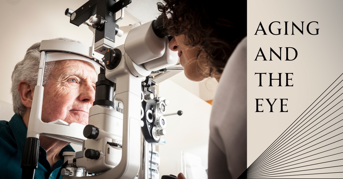
Have you ever wondered what happens to the visual system as we age? What does the term "second sight" mean? What is presbyopia? What are the eyes more susceptible to as the aging process occurs? What can be done to prevent certain aging factors of the eye? The answer lies in a theory known as apoptosis (no that's not the name of the latest pop artist).
Apoptosis is the pre-programmed life of every cell in our body. Most studies show that it's a function of our programmed DNA. It's the ability for cells to survive and thrive in the anatomical environment. The body's ability to withstand and thrive during the aging process depends on proper nutrition, good mental health, exercise, and adequate oxygen supply. That's why studies have shown smoking can shorten your life by a decade or more.
In regards to aging and the eye, there is a phenomenon during the 6th to 7th decade of life called "second sight." This is simply progressive nearsightedness in older adults secondary to cataracts. Close to 50% of the population over 60 years old has cataracts. Cataracts are a clouding of the natural lens of the eye that can impair vision, causing glare and loss of detail. When patients experience second sight, it is sometimes quite convenient for them--they see up close without the reading glasses they have been depended on since their 40s.
Another aspect of the aging process is losing the reading vision you had all your life. This is called Presbyopia. Presbyopia is a Latin term which means "old eyes."
What happens in Presbyopia?
Before our mid-forties, the natural lens of the eye is very pliable and can easily focus on items up close. But in our mid-forties, the lens tends to lose its elasticity. When experiencing presbyopia, people generally hold reading material farther away to see it more clearly. Presbyopia can be managed through bifocal or multifocal glasses or contact lenses, and some surgeries.
As aging occurs, the eyes are more susceptible to cataracts, glaucoma, macular degeneration and vascular disorders of the eye as well as dry eye syndrome.
To help prevent and manage these conditions, there are a variety of options.
- Maintaining yearly dilated eye exams for preventative care.
- Protect your eyes against the sun with UV sunglasses.
- Take antioxidant vitamins to help bolster the protection of the macula.
- Use artificial tears to hydrate the eye and keep your body hydrated by drinking plenty of water.
- Keep emotional, physical, and mental stress to a minimum.
Being educated on how we age is the first step towards good ocular health and diminished chances of early apoptosis.
The content of this blog cannot be reproduced or duplicated without the express written consent of Eye IQ.
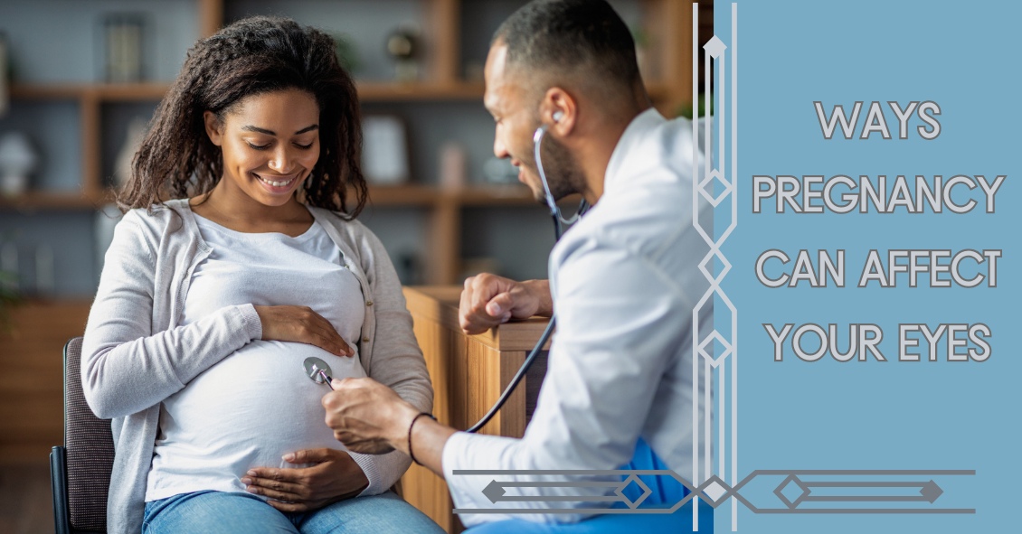
We all know that during pregnancy, a woman's body goes through a great deal of change hormonally and physiologically. But did you know her eyes change as well? Below are some of the most common effects pregnancy can have on the eye.
- Corneal changes. In some cases, pregnancy can cause the cornea, the front window of the eye, to change curvature and even swell, leading to shifts in glasses and contact lens prescriptions. In addition, changes in the chemistry of the tear film can lead to dry eyes and contact lens intolerance. It is for these reasons that it is generally not recommended to have any new contact lens fitting or new glasses prescription checks until several months postpartum. We want to get the most accurate measurements possible.
- Retinal changes. Many different conditions can affect the retina during pregnancy. If the pregnant woman has diabetes, diabetic eye disease can progress by 50%. In women with preeclampsia, a condition where blood pressure rises significantly, over 40% of women can show changes in the retinal blood vessels, and 25% to 50% complain of changes to their vision.
- Eye Pressure Fluctuation. Intraocular pressure (IOP) usually decreases during pregnancy. The exact mechanism causing this is unknown, but it is usually attributed to an increase of flow of intraocular fluid out of the eye. This is good news for pregnant women with glaucoma or high IOP. In fact, the drop in IOP is larger when you start with a high IOP compared to one in the normal range.
There are many more effects that pregnancy can have on the eye, but these are the most common. One other thing to keep in mind is that though the likelihood of any adverse effect is extremely low, we try not to use any diagnostic eye drops on pregnant patients during the eye exam. Unless there is a medical necessity to dilate the pupils or check IOP, it is a good rule of thumb to put off using drops until after the patient has given birth in order to protect the developing baby.
Article contributed by Dr. Jonathan Gerard
This blog provides general information and discussion about eye health and related subjects. The words and other content provided in this blog, and in any linked materials, are not intended and should not be construed as medical advice. If the reader or any other person has a medical concern, he or she should consult with an appropriately licensed physician. The content of this blog cannot be reproduced or duplicated without the express written consent of Eye IQ.
1. Vision is so important to humans that almost half of your brain’s capacity is dedicated to visual perception.
2. The most active muscles in your body are the muscles that move your eyes.
3. The surface tissue of your cornea (the epithelium) is one of the quickest-healing tissues in your body. The entire corneal surface can turn over every 7 days.
4. Your eyes can get sunburned. It is called photokeratitis and it can make the corneal epithelium slough off just like your skin peels after a sunburn.
5. Ommatophobia is the fear of eyes.
6. You blink on average about 15 to 20 times per minute. That blink rate may decrease by 50% when you are doing a visually demanding task like reading or working on a computer – and that’s one reason those tasks can lead to more dry-eye symptoms.
7. Your retinas see the world upside down, but your brain flips the image around for you.
8. If you are farsighted (hyperopia) your eye is short, and if you are shortsighted (myopia) your eye is long.
9. An eyelash has a lifespan of about 5 months. If an eyelash falls out it takes about 6 weeks to fully grow back.
10. One in every 12 males has some degree of “color blindness.”
Article contributed by Dr. Brian Wnorowski, M.D.
This blog provides general information and discussion about eye health and related subjects. The words and other content provided on this blog, and in any linked materials, are not intended and should not be construed as medical advice. If the reader or any other person has a medical concern, he or she should consult with an appropriately licensed physician. The content of this blog cannot be reproduced or duplicated without the express written consent of Eye IQ.